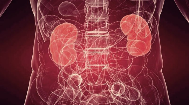
Kidneys are well-protected organs located in the retroperitoneum or the posterior/rearmost part of the abdomen, just below the diaphragm. They are surrounded by fat and are mobile due to movements of the diaphragm during respiration. This movement is limited by short blood vessels connected to the kidneys. In some people who are very thin, due to lack of fat around the kidneys, the kidneys descend and rotate anteriorly, resulting in what is called ‘renal ptosis’, ‘falling kidney’, ‘nephroptosis’ or ‘floating kidney’.
Floating kidney
This rare condition presents usually with pain over the upper abdomen and flank. It is more common on the right side. The exact origin of the symptoms is not known but is thought to be due to obstruction of the blood supply of the kidneys or of the outflow of urine due to rotation of the kidney. The obstruction to the outflow of urine results in ‘hydronephrosis’ or distension of the kidney with urine. This causes stretching of the outer layer or capsule resulting in pain. Obstruction of blood supply causes kidney ischemia (reduced oxygen supply through the blood) and pain. The pain can also be due to the stretching of nerves around the blood vessels.
The typical patient is a young (20 – 40-year-old), a thin female who complains about pain in the erect/upright posture or while walking. The pain is relieved by upward movement of the kidney which is achieved by lying down on the bed with legs elevated on pillows or in the knee-chest position. The most severe presentation is called ‘Dietl’s crisis’, which manifests with severe cramp-like abdominal pain, nausea, vomiting, fever/chills, increased heart rate, and reduced amount of urine. Patients may also develop transient hematuria (blood loss in the urine) or proteinuria (loss of proteins in the urine). Some patients may also develop chronic urinary tract infections. Long-term complications include hypertension and kidney stones.
Diagnosis
On imaging by computerized scanning or x-rays (Intravenous pyelogram), the kidney is found to descend more than the length of two vertebral bodies or more than 5 cm on change of posture from lying down position to upright standing position.
Nuclear imaging can also be used to demonstrate the obstruction of blood flow or the urine outflow in an erect posture.
Colour Doppler Flow Imaging (CDFI) can be used to demonstrate a difference in blood flow in the blood vessels of the kidney in the upright position of the patient. In this posture, the blood flow to the affected kidney is reduced. On lying down the flow pattern returns to normal.
Treatment of floating kidney
The treatment for a floating kidney is done by surgery in symptomatic patients. It takes about one to two hours to complete this procedure. It is called ‘nephropexy’ which means ‘fixing of the kidney’. It can be done by open surgery or by the keyhole/laparoscopic method. The laparoscopic approach is preferred because of lesser postoperative pain, better cosmetic results, and early return to work.
Laparoscopic Nephropexy is done through a minimally invasive approach. The kidney is completely exposed and then the outer layer of the capsule is stitched to the surrounding muscles (fascia overlying psoas and quadratus lumborum muscles). Other methods of fixing may use metal clips, polyglactin mesh, or adhesive tissue glue. In the studies conducted in the past, it has been found significant improvement in kidney function after surgery. Another study demonstrated a 91% improvement in the pain symptoms.
Circle (U) nephrostomy tube insertion – in this surgical method, a ‘U’ shaped tube is passed through the kidney so as to hook it up to the 12th rib (lowermost rib). This tube is removed after 2 to 3 weeks during which scar forms between the kidney and rear abdominal wall. This fixes the kidney and prevents its abnormal mobility.
The complications include recurrence of symptoms due to failure of surgery when the patient develops ptosis/falling of kidney.
The floating kidney is a rare condition. If you are very thin or have recently lost weight, and have pain abdomen while walking or running, it would be good to consult your doctor. If undetected, then chronic infections or obstructions may result in permanent loss of kidney function.