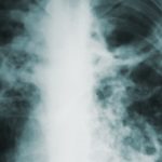
Croupous pneumonia
 Croupous pneumonia – refers to common diseases. 6-18 cases per 1 thousand population per year, such is the prevalence of this type of pneumonia among the population of different countries. As a rule, it becomes more frequent during a flu epidemic.
Croupous pneumonia can develop at any age, but children are more likely to get sick, in the elderly the disease is especially difficult. Adult men suffer from this disease almost 3 times more often than women.
Etiology
Over the past 10-15 years, croupous pneumonia has fundamentally changed its clinical course. One of the reasons for this polymorphism is a change in the causative agent of the disease.
If previously pneumococcus was considered to be almost the only causative agent of pneumonia, which has not lost its significance today, now associations of microorganisms, including viral and bacterial ones, such as Friedlander diplobacilli, Pfeifer’s bacillus, streptococci, play a significant role.
However, for the occurrence of the disease, the presence of microbes in the body or the ingestion of it is not enough. Often they are in the body, in particular in the oral cavity and upper respiratory tract, without causing disease. As S. S. Zimnitsky said, lobar pneumonia is “a contagious disease, but not contagious.” It is little contagious as it is caused not only by the introduction of pneumococcus into the upper respiratory tract of a person, but also by various predisposing factors. For example, cooling, reduced body resistance, injuries to the chest and skull, or nervous shocks, alcohol and physical fatigue can contribute to the onset of the disease. The sensitization of the body and its increased sensitivity to infections are also important.
Pathogenesis
Pathogens enter the lung tissue by the bronchogenic, hematogenous and lymphogenous pathways. Most often, pneumococci penetrate through the bronchi and invade the lungs, starting from the root area, where they settle in the lymphatic vessels, causing inflammation, first of the interstitial tissue, and then of the interalveolar septum. From here, the infection enters the lumen of the alveoli, which leads to the formation of a fibrinous effusion. As a result, the lung becomes dense, airless, hepatization occurs. During this period, the affected lung contains large quantities of virulent pneumococci, which are excreted in sputum and are found in the blood, and antibody formation occurs here simultaneously. When the antibody titer reaches a certain level, pneumococci die and disappear from sputum and blood. In the lung tissue, the release of the proteolytic enzyme and the resorption of fibrinous effusion are enhanced. An important role in the pathogenesis of croupous pneumonia is played by violations of the protective mechanisms of the bronchopulmonary system and the state of humoral and tissue immunity. The survival of bacteria in the lungs, their reproduction and distribution along the alveoli largely depends on their aspiration with mucus from the upper respiratory tract and bronchi – which is favored by cooling, from the excessive formation of edematous fluid, covering an entire fraction or several lobes of the lungs. At the same time, immunological damage and inflammation of the lung tissue due to a reaction to the antigenic material of microorganisms and other allergens is possible.
Pathological anatomy
In pathological anatomy with typical croupous pneumonia, there are 4 stages of changes in the lung tissue:
- Stage of the tide, in which there is hyperemia and inflammatory edema of the lung tissue with the accumulation in the alveoli of liquid serous exudate with an admixture of red blood cells, white blood cells. The exudate contains pneumococci and fibrin fibers. This stage lasts from 12 hours to 3 days.
- The stage of red hepatization is characterized by compaction of fibrin in the exudate with an abundant admixture of red blood cells and a small amount of leukocytes. The lung is macroscopically compacted and enlarged. The duration of this stage is 1-3 days.
- Stage gray hepatitis is not hyperemia. She noted the disappearance of red blood cells from the exudate with a simultaneous increase in the number of leukocytes in it, which gives the affected part of the lungs a gray-green color. The inflamed lobe of the lung is still compacted, enlarged, the alveoli are filled with a dense mass of fibrin.
- Stage resolution, during which dissolution and resorption of fibrin by proteolytic enzymes occurs, the breakdown of leukocytes is observed. The exudate becomes fluid and purulent. There comes its complete resorption, in which the lung becomes soft. This stage is the longest, its duration depends on the prevalence of the process, the reactive characteristics of the body, etc.
Along with lung damage in the pleura, changes occur in the form of fibrinous overlays on the pleural surface of the affected lung. Due to the spread of infection along the lymphatic tract, bronchial lymph nodes increase.


