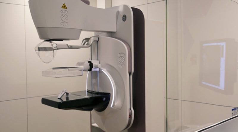
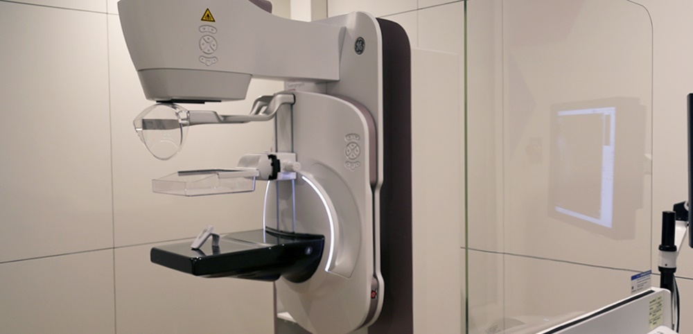
Mammography is an imaging modality in which radiographs of breasts are taken to detect changes suggestive of cancer. It uses a low dose of x-rays for visualization of any structural changes in the architecture of breast parenchyma.
Types of Mammography
Mammography is basically of two types depending on the indication for which it is employed. It is classified as screening mammography and diagnostic mammography.
Screening mammography: Screening mammography is done in asymptomatic women for the detection of breast cancer. Screening investigations are done in a population where the disease is commonly found. This helps in the early detection of disease before it progresses to become symptomatic. A clinical study demonstrated that screening mammography resulted in a 33% reduction in mortality in women. Mammography was found to be more accurate than clinical examination for the detection of early breast cancers. In this investigation, two views of each breast are taken. One is craniocaudal (in the vertical axis of the breast) and the other is a mediolateral oblique view (along the horizontal axis of the breast). With these two views, a maximum amount of breast tissue can be imaged. It delivers a radiation dose of 0.1 centigrade and it does not result in an increased risk of breast cancer.
Types of mammography and mammography for detecting breast cancer
The NCCN (National Comprehensive Cancer Network) suggests that, from the age of 40 years, yearly screening mammography should be done. Studies of mammography screening confirm a 40% reduction in invasive and advanced stage (stage II, III, and IV) of breast cancer and a 30% increase in overall survival.
Diagnostic mammography: This imaging modality is used for the assessment of breast in women who present with breast symptoms suspicious of cancer. They include a lump, nipple discharge, nipple retraction, ulcer over breast which fails to heal, enlargement of the breast, and a lump in the axilla. Imaging of both breasts is done. It helps in finding out a bilateral (involving both breasts) disease. It may also show evidence of multifocality (multiple areas of cancer in a quadrant of the breast) or multicentric disease (involving two or more quadrants of the breast).
It involves taking the two views as described in screening mammography; the craniocaudal and mediolateral oblique views. If there is a suspicious area, additional images like 90 degree lateral and spot compression views can be taken. The compression view reduces the amount of tissue between the imaging sensor and the tumor and gives a better-resolution image with a lower radiation dose. Magnified views may also be obtained to see the margins of a lump or small calcifications. These views help in the identification of the exact location of the tumor in the breast. This information can be used to guide needle localization and needle biopsy of tumors.
Mammography in breast cancer detection
The mammographic features which are indicative of cancer include a mass with irregular margins, asymmetric thickening of tissue inside the breast, and clustered deposits of micro-calcifications. The presence of micro-calcifications is associated with 50% of breast cancers in which no lump or mass is palpable. These are especially important in young patients in whom these micro-calcifications are the only findings on mammography.
Xeromammography: This technique is similar to conventional mammography except that the image is recorded on a xerography plate (like the photographic paper) which provides a positive image. In mammography, we get a negative image.
Digital mammography uses a digital CCD sensor to record the image. It also allows manipulation of the contrast in the image which results in better visualization of suspicious areas. It is especially useful in women with large breasts, dense breasts, and young women less than 50 years of age.
Mammography is an important breast imaging modality that uses a low dose of x-rays for the early detection of breast cancer. It has a great role to play in deciding the type of treatment to be offered to these patients. It has resulted in improving the overall survival rate of patients with breast cancer.